-
 Epithermal deposit
-
 Rhizoid
-
 Fanconi anaemia
-
 ATPase
-
 Mean coordinates
-
 Cathode
-
 CDP
CDP
-
 Screening
-
 Open cluster
-
 Phytochrome
-
 Tadelakt
-
 Newton
-
 PP Live
-
 Monophyletic
-
 mRNA
mRNA
-
 Hydrogen bond
-
 CD-Photo
-
 Principle of complementarity
-
 Bioenergy
-
 Reverse genetics
-
 Holohedry
-
 Micral
-
 Montrachet
-
 NASA World Wind
-
 Crookes tube
-
 ISP
ISP
-
 Cranberry
-
 SNMP
SNMP
-
 Cleavage
-
 Genu valgum
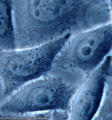
Phase contrast microscope
A phase contrast microscope has been used for a very long time.
A phase contrast. © polymtl.ca
microscope Technique of
Contrast microscopy
A phase contrast microscope (is a light microscope) which converts differences in refractory index between two structures into contrast levels which result in differences in phase for light waves which pass through them. It therefore visualises transparent structures when their refractory index is different from the neighbouring refractory index.
Two devices known as phase rings are positioned, one in the condenser (the optical system which focuses light on the object) and the other in the lens of this type of microscope. When the edge of a structure produces sufficient diffraction, the light crossing through it changes phase compared to the other light rays. The rings filter these different phase rays producing an accentuated contrast image of the structure. It was designed in 1930 by the Dutchman, Frits Zernike (1888-1966), and won its designer the Nobel Prize for Physics in 1953.
Use
This microscope allows living cells to be studied without needing to stain them. The phase contrast microscope produces a halo around the structure seen, which does not happen with an interference contrast microscope.
 Observation of cells by a phase contrast microscope. © Exothermic, CC by-nc-sa 2.0
Latest
Fill out my online form.


