-
 Electrolyte
-
 Smart phone
-
 Demersal
-
 Motility
-
 Metastasis
-
 Scots pine
-
 Up quark
-
 Addiction
-
 Central antihypertensive
-
 Western red cedar
-
 Vitelline sac
-
 Effinergie
-
 Erenna
-
 Accretionary prism
-
 Necrosis
-
 Constellation of Andromeda
-
 Poisson brackets
-
 ID3
ID3
-
 Heterotrophism
-
 Slurry pipeline
-
 Human papillomavirus
-
 Elongated facies
-
 Debye force
-
 Weightlessness
-
 Swallowtail
-
 Empress tree
-
 Galileo
-
 Lychee
-
 Antibiotic resistance
-
 Biopiracy
Transmission electron microscope
A transmission electron microscope is an electron microscope used to see objects far smaller than cells.
Transmission electron microscopetechnique
The transmission electron microscope (TEM) uses a high voltage electron beam emitted by an electron gun. Electromagnetic lenses are used to focus the electron beam on the sample. As the electron beam passes through the sample and the atoms it is composed of, it produces different forms of irradiation. In general, only transmitted electrons are then analysed by the detector that translates the signal into a contrast image.
Samples must be prepared using a specific protocol which must both preserve their structure and make it a conductor to allow the electron beam to pass through. Very thin sample sections are prepared using an ultramicrotome (60 to 100 nanometres). Heavy metal stains can also be used to increase the specific structures in the samples that have been placed on the observation grids.
Using of the transmission electron microscope
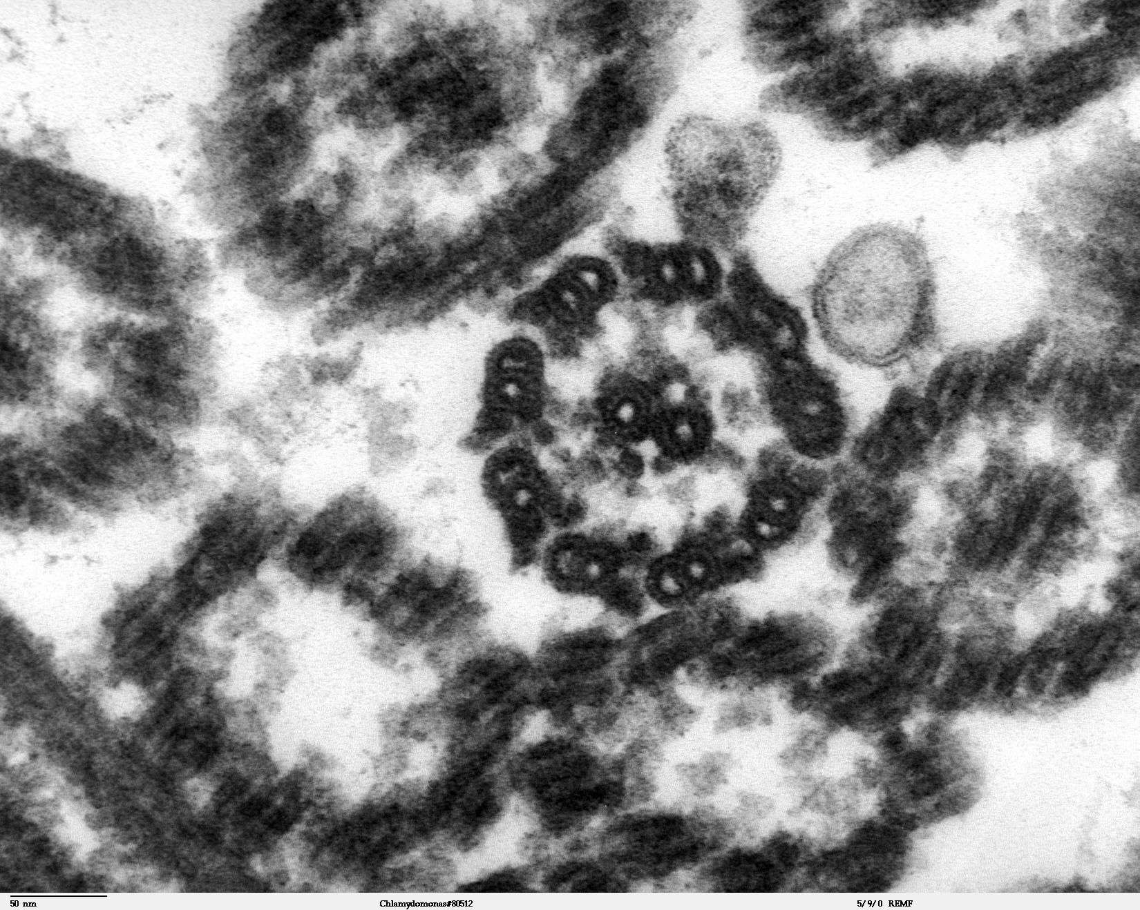
While sample preparation takes longer and is more demanding than for light microscopy, the resolution offers an unparalleled view of structures. The transmission electron microscope can visualise intracellular organelles, viruses, and crystals, etc.
 A section of a Chlamydomanas reinhardtii flagellum seen under transmission electron microscopy. © DR
Latest
Fill out my online form.


